



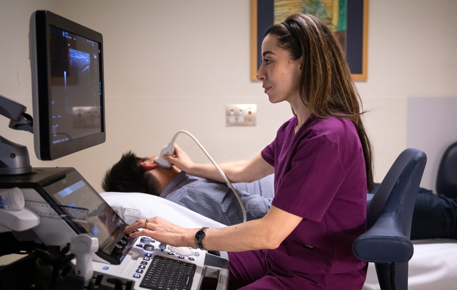
Ultrasound scans offer real-time images of your body’s interior using sound waves. Unlike x-rays, ultrasounds do not involve radiation exposure.
We direct high-frequency sound waves at the body part examined during the ultrasound. These sound waves are emitted by a device called a transducer, which also captures the echoes that bounce back from your body. The reflected sound waves, or echoes, generate an image displayed on a monitor, allowing your healthcare team to visualise the inside of your body in real-time.
Our ultrasound specialists at St Vincent’s Private Radiology are ideal for completing abdominal, vascular, obstetrics and musculoskeletal studies. For gynecological studies, you may require an internal examination that provides greater structural detail. This will be discussed with you prior to the examination.

A Sonographer utilises a hand-held scanner, called a transducer to perform an ultrasound. High-frequency sound waves are sent into your body, and their echoes are converted into electrical impulses to create images on a screen.
During the scan, the sonographer will apply a gel to your skin in the examined area. The sonographer moves the transducer on the gel, pressing gently if necessary.
Ultrasound scans take 20-60 minutes and are performed by specially trained professionals. There are no after-effects, and you can resume your normal activities afterwards.


All diagnostic imaging reports are sent directly to your referring healthcare provider.
Access to reports via MyHealth Record is currently unavailable while we complete the required integration. We are working to install this service and will update our website once this has been completed.

We are committed to providing affordable competitive out of pocket fees.
For Medicare rebatable examinations, We Bulk Bill Pension or Concession card holders and full-time tertiary students.
If you have any questions regarding fees or billing, please contact our administration team.