



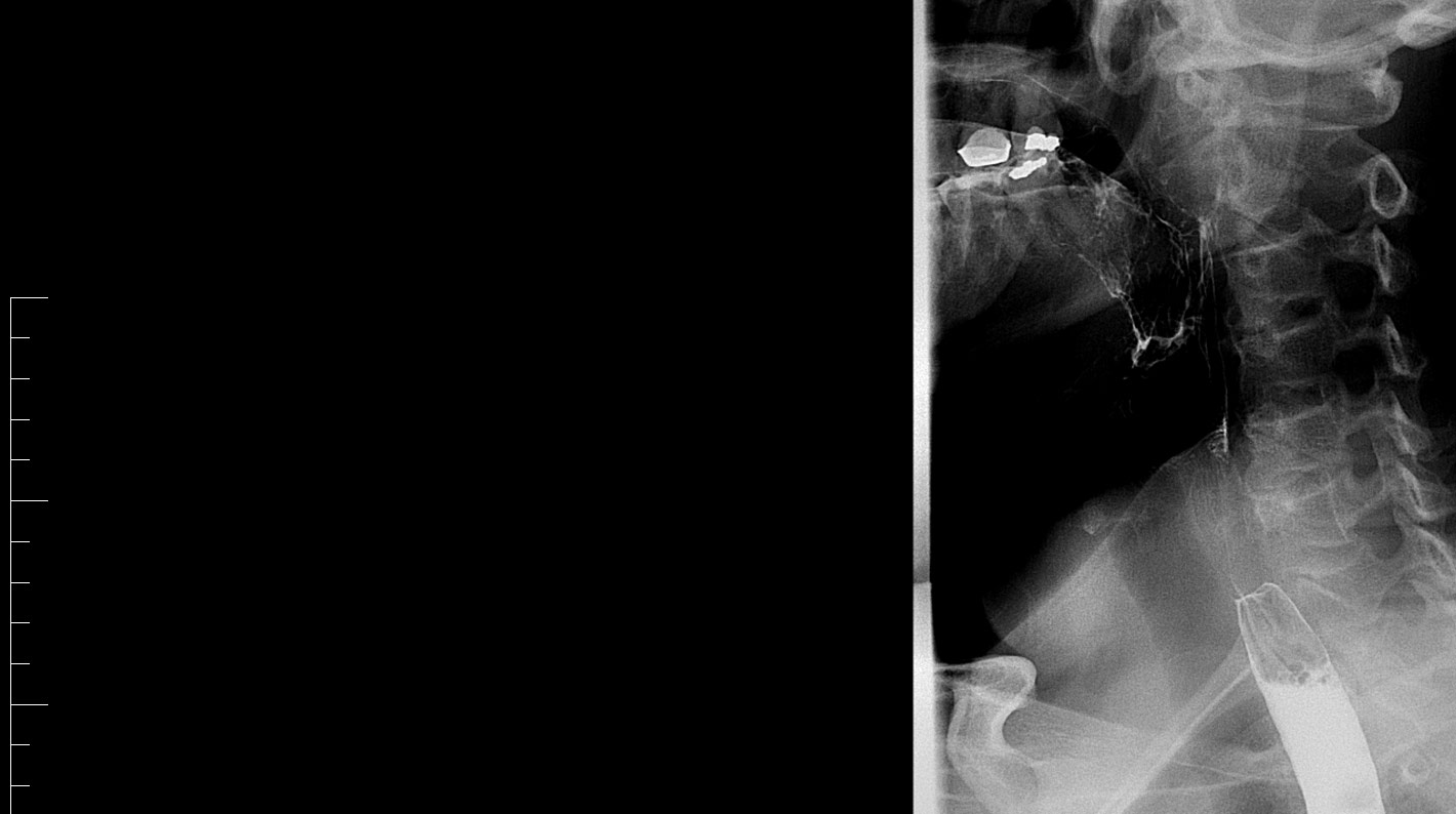
Fluoroscopy is a medical imaging technique that helps doctors examine the inside of your body in real-time. It uses x-rays to create live, moving images, allowing healthcare professionals to see how your organs function and observe any abnormalities. Unlike traditional x-rays, which capture motionless pictures, fluoroscopy captures a continuous series of images, like a video.
During a fluoroscopy procedure, you may be asked to eat or drink a contrast material. This substance helps improve the visibility of specific organs or areas in your body, providing a clearer, more detailed image for your doctor to analyse.
Fluoroscopy is used for diagnosing and treating conditions related to the digestive system, blood vessels, joints, or other organs.

Depending on the specific exam, you might be asked to fast, or consume a contrast material to enhance the images. You may be asked to change into a hospital gown and remove any jewellery or metal objects.
During our examination, you will be positioned on an examination table, and the x-ray machine will be positioned above or beside you. The machine captures live images, allowing your treating team to observe your body’s internal structures in motion.


All diagnostic imaging reports are sent directly to your referring healthcare provider.
Access to reports via MyHealth Record is currently unavailable while we complete the required integration. We are working to install this service and will update our website once this has been completed.

We are committed to providing affordable competitive out of pocket fees.
For Medicare rebatable examinations, We Bulk Bill Pension or Concession card holders and full-time tertiary students.
If you have any questions regarding fees or billing, please contact our administration team.